Which diagnosis is most likely confirmed by echocardiography in a 65-year-old female presenting with new onset chest pain associated with ST segment elevation on the electrocardiogram and angiographically normal coronary artenes?
Correct Answer:
D
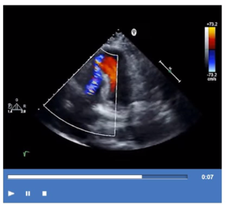
Takotsubo cardiomyopathy, also known as stress-induced cardiomyopathy or 'broken heart syndrome,' predominantly affects postmenopausal women (usually older than 50 years) and often presents with acute chest pain and ST-segment elevation on the ECG mimicking acute myocardial infarction. However, coronary angiography reveals normal or non-obstructive coronary arteries.
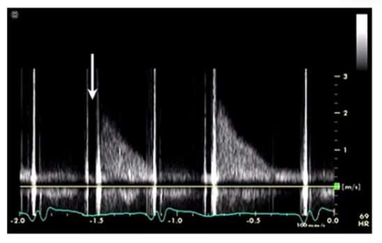
Echocardiographically, Takotsubo cardiomyopathy is characterized by transient left ventricular systolic dysfunction with a typical pattern of apical ballooning and basal hyperkinesis. The wall motion abnormality extends beyond a single coronary artery territory, differentiating it from ischemic cardiomyopathy.
The diagnosis is supported by the clinical presentation, typical echocardiographic findings, and exclusion of obstructive coronary artery disease. The condition is usually reversible over days to weeks.
This is extensively described in the 'Textbook of Clinical Echocardiography, 6e' (Chapter 8: Coronary Artery Disease and Takotsubo Syndrome), which highlights the typical patient demographics, presentation, echocardiographic features, and prognosis .