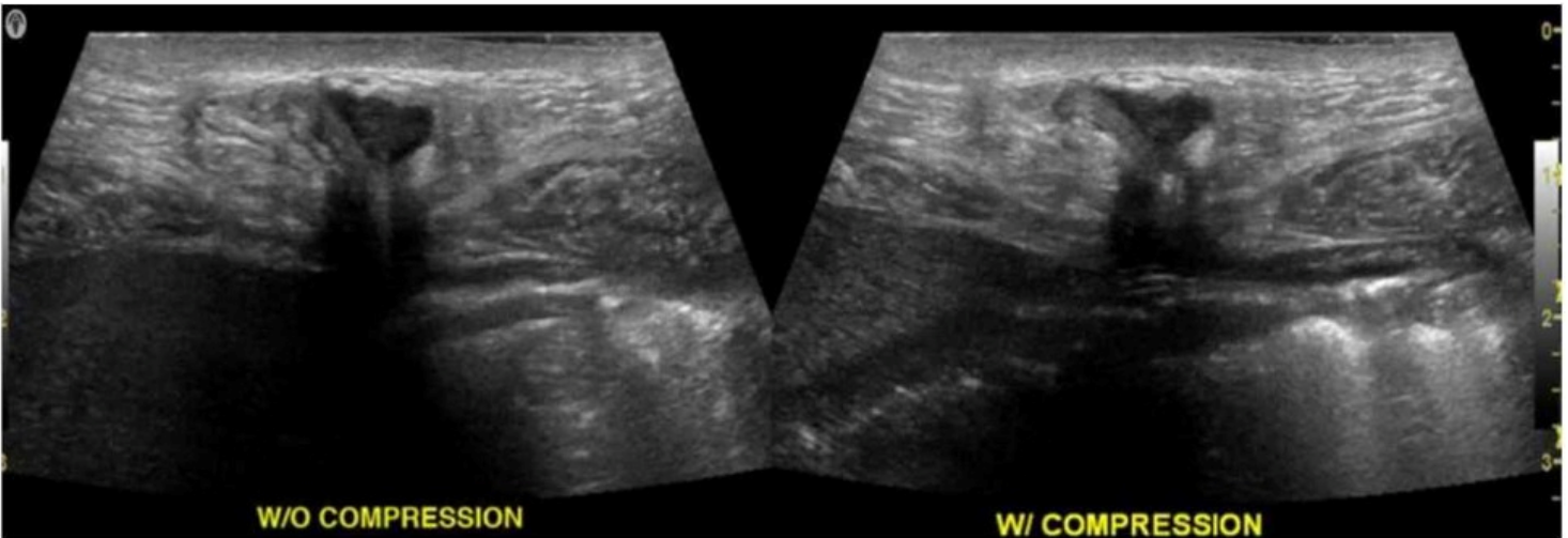
ARDMS AB-Abdomen Exam Dumps

Get All Abdomen Sonography Examination Exam Questions with Validated Answers

AB-Abdomen Pack
Vendor: ARDMS
Exam Code: AB-Abdomen
Exam Name: Abdomen Sonography Examination
Exam Questions: 165
Last Updated: January 7, 2026
Related Certifications: Registered Diagnostic Medical Sonographer
Exam Tags: Professional ARDMS abdominal sonographers and medical sonographers
Gurantee
  • 24/7 customer support
  • Unlimited Downloads
  • 90 Days Free Updates
  • 10,000+ Satisfied Customers
  • 100% Refund Policy
  • Instantly Available for Download after Purchase

Get Full Access to ARDMS AB-Abdomen questions & answers in the format that suits you best

PDF Version

$40.00
$24.00
  • 165 Actual Exam Questions
  • Compatible with all Devices
  • Printable Format
  • No Download Limits
  • 90 Days Free Updates

Discount Offer (Bundle pack)

$80.00
$48.00
  • Discount Offer
  • 165 Actual Exam Questions
  • Both PDF & Online Practice Test
  • Free 90 Days Updates
  • No Download Limits
  • No Practice Limits
  • 24/7 Customer Support

Online Practice Test

$30.00
$18.00
  • 165 Actual Exam Questions
  • Actual Exam Environment
  • 90 Days Free Updates
  • Browser Based Software
  • Compatibility:
    supported Browsers

Pass Your ARDMS AB-Abdomen Certification Exam Easily!

Looking for a hassle-free way to pass the ARDMS Abdomen Sonography Examination exam? DumpsProvider provides the most reliable Dumps Questions and Answers, designed by ARDMS certified experts to help you succeed in record time. Available in both PDF and Online Practice Test formats, our study materials cover every major exam topic, making it possible for you to pass potentially within just one day!

DumpsProvider is a leading provider of high-quality exam dumps, trusted by professionals worldwide. Our ARDMS AB-Abdomen exam questions give you the knowledge and confidence needed to succeed on the first attempt.

Train with our ARDMS AB-Abdomen exam practice tests, which simulate the actual exam environment. This real-test experience helps you get familiar with the format and timing of the exam, ensuring you're 100% prepared for exam day.

Your success is our commitment! That's why DumpsProvider offers a 100% money-back guarantee. If you don’t pass the ARDMS AB-Abdomen exam, we’ll refund your payment within 24 hours no questions asked.
 

Why Choose DumpsProvider for Your ARDMS AB-Abdomen Exam Prep?

  • Verified & Up-to-Date Materials: Our ARDMS experts carefully craft every question to match the latest ARDMS exam topics.
  • Free 90-Day Updates: Stay ahead with free updates for three months to keep your questions & answers up to date.
  • 24/7 Customer Support: Get instant help via live chat or email whenever you have questions about our ARDMS AB-Abdomen exam dumps.

Don’t waste time with unreliable exam prep resources. Get started with DumpsProvider’s ARDMS AB-Abdomen exam dumps today and achieve your certification effortlessly!

Free ARDMS AB-Abdomen Exam Actual Questions

Question No. 1

Which change of the inferior vena cava spectral Doppler waveform is expected superior to a nonocclusive thrombus?

Show Answer Hide Answer
Correct Answer: C

In the presence of a nonocclusive thrombus, Doppler waveform above the thrombus typically shows dampened flow with loss of normal respiratory phasicity due to partial venous outflow obstruction. Complete absence of flow is typically seen with occlusive thrombus.

According to Zwiebel's Introduction to Vascular Ultrasound:

''Partial obstruction produces dampened and continuous flow patterns superior to a nonocclusive thrombus.''


Zwiebel WJ, Pellerito JS. Introduction to Vascular Ultrasound. 6th ed. Elsevier, 2019.

AIUM Practice Parameter for Venous Ultrasound, 2020.

---

Question No. 2

Which sonographic appearance of the normal epididymis is the most common?

Show Answer Hide Answer
Correct Answer: C

The normal epididymis typically appears as a homogeneous structure that is either isoechoic or slightly hypoechoic compared to the testis. The most accurate description is 'homogeneous compared to the testis,' meaning the texture is uniform. It is not anechoic, nor does it typically show irregular borders unless pathology is present.

According to Rumack's Diagnostic Ultrasound:

'The normal epididymis appears homogeneous and is isoechoic or slightly hypoechoic relative to the testis.' (Rumack CM et al., Diagnostic Ultrasound, 5th ed.)


Rumack CM, Wilson SR, Charboneau JW, Levine D. Diagnostic Ultrasound. 5th ed. Elsevier; 2017.

AIUM Practice Parameter for Scrotal Ultrasound, 2020.

---

Question No. 3

Which exam type would be most beneficial for evaluating an elderly male patient who is complaining of dysuria and nocturia?

Show Answer Hide Answer
Correct Answer: A

In elderly men, dysuria and nocturia often suggest lower urinary tract symptoms such as benign prostatic hyperplasia (BPH). A pelvis limited ultrasound is most appropriate to evaluate the bladder and prostate for enlargement, residual urine volume, or other causes of obstructive symptoms.

According to AIUM Practice Guidelines:

''Pelvic ultrasound can evaluate bladder volume, post-void residual, and prostate size in men with lower urinary tract symptoms.''


AIUM Practice Parameter for the Performance of Ultrasound of the Prostate, 2020.

Rumack CM, Diagnostic Ultrasound, 5th ed. Elsevier, 2017.

---

Question No. 4

Which finding is indicated by the arrow in this image of the right upper quadrant?

Show Answer Hide Answer
Correct Answer: B

The image provided is a right upper quadrant (RUQ) ultrasound---typically performed during a FAST (Focused Assessment with Sonography in Trauma) exam or for abdominal assessment. The arrow points to an anechoic (black) fluid collection seen above the diaphragm and posterior to the liver.

This fluid collection lies within the thoracic cavity, confirming the diagnosis of a pleural effusion. Pleural effusions are seen sonographically as an anechoic or hypoechoic area superior to the diaphragm in the thoracic cavity and often appear triangular or crescent-shaped. The diaphragm is visualized as a curvilinear echogenic structure separating the liver (or spleen) below from the lung space above.

Comparison of answer choices:

A . Retroperitoneal hemorrhage would be seen in the posterior abdomen, not above the diaphragm.

B . Pleural effusion is correct---anechoic fluid above the diaphragm is classic for this condition.

C . Mirror image artifact occurs when liver echoes are mirrored across the diaphragm and lung---this is not a mirror artifact.

D . Ascites collects inferior to the diaphragm and around the abdominal organs, not in the thoracic cavity.


Ma OJ, Mateer JR, Blaivas M. Emergency Ultrasound, 3rd ed. McGraw-Hill; 2014.

Moore CL, Copel JA. Point-of-care ultrasonography. N Engl J Med. 2011;364(8):749--757.

Rumack CM, Wilson SR, Charboneau JW, Levine D. Diagnostic Ultrasound, 5th ed. Elsevier; 2017.

Question No. 5

Which technique best differentiates a bladder mass from a hematoma?

Show Answer Hide Answer
Correct Answer: D

Changing the patient's position allows evaluation of lesion mobility. Blood clots and hematomas are often mobile, while true bladder wall masses remain fixed. This technique helps differentiate between solid masses and non-adherent debris.

According to Rumack's Diagnostic Ultrasound:

''Changing patient position may distinguish between mobile blood clots and fixed bladder wall masses.''


Rumack CM, Wilson SR, Charboneau JW, Levine D. Diagnostic Ultrasound. 5th ed. Elsevier, 2017.

AIUM Practice Parameter for Bladder Ultrasound, 2020.

100%

Security & Privacy

10000+

Satisfied Customers

24/7

Committed Service

100%

Money Back Guranteed